Physics Research

Vision and ambitions
Our mission is to develop methods to enable precise irradiation of tumors while sparing normal tissues.
The combination of physics and artificial intelligence offers opportunities to develop new methods to improve several steps in the radiotherapy process.
Our ambition is to be a leader in the development of technology, both for preclinical fundamental cancer research, and for treatment of patients.
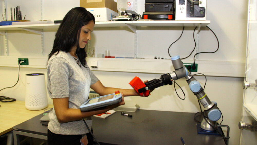
In brachytherapy we improve dose delivery by developing advanced (Monte Carlo) dose calculations, verification and imaging, as well as novel applicators. Maastro is pushing the boundaries in brachytherapy within measurement of accuracy and precision, and safety of treatments
The aim of DGRT is accurate measurement of the true dose delivered to the patient. Artificial intelligence methods are used to create decision models for adaptive radiotherapy using DGRT.
This research line concerns a novel imaging technique, to obtain more detailed information about patient anatomy and tissue compositions, which is needed for accurate dose calculations. Research is performed on imaging and dose calculation accuracy, as well as on automatic contouring.
Our proton radiotherapy clinic has a synchrocyclotron (Mevion) for proton therapy, with advanced beam shaping capabilities, real-time patient tracking system, and integrated dual-energy CT imaging.
We developed a novel research platform for research with small animals. With an integrated image guidance and precision irradiation system with millimeter-size beams, we can use x-ray CT, fluoroscopy and bioluminescence imaging.
We combine the latest artificial intelligence methods to automatically contour organs, to speed up dose calculations, to reconstruct images, and to detect treatment errors.


